Atopy (literally "unusualness") is a medical collective term for different kinds of hypersensitivity. One of these is atopic dermatitis (AD), an allergic skin disease in humans and animals. About 10-15% of domestic dogs suffer from this disorder. Mainly affected are dogs from the age of 6 months up to 3 years.
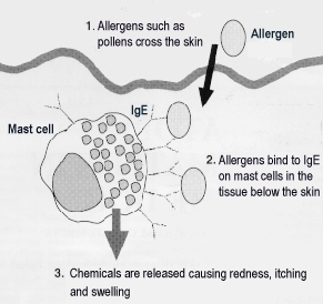 The cause and development of this disease has not yet been completely clarified. In dogs the organism generates antibodies of the type IgE (immuglobulin E) against normally harmless substances such as house dust mites, pollen, grasses, insects, animal hair and others. The allergy-causing substance, the allergen, is absorbed through the skin (percutaneously). The allergens bind to copiously produced IgE antibodies which coat specific defence cells (mast cells). The mast cells thereupon release chemical substances causing itching, redness and swelling. The most common allergens are house dust mites and pollen.
For a long time a polygenic inheritance (dependent on several genes) was assumed in AD. Recently however there has been evidence of a mutated cell or a special "atopy-gene" as the sole origin.
Symptoms
The symptoms of atopic dermatitis are itching (pruritus) and redness of the skin (erythema). A first sign is itching without skin alteration, after this primary skin lesions with redness follow.
Itching affects the face (eyes, chin, flews), ears and paws, axillary, ventral and inguinal regions, elbows, metacarpals and ankle joints. In some dogs also the anal region is affected. Least of all the back is affected, if it is affected at all.
Soon in the further course secondary skin lesions appear caused by scraping, licking (discolouration of the coat) or gnawing at the itchy sections of the skin. These secondary skin lesions are inflammation with bacteria and/or yeast fungus with their typical symptoms (pustules, scabs, hair loss), increased development of dandruff and finally chronic skin lesions with baldness, thickening and black colouring of the skin, rancid smell etc.
Diagnosis
If an allergic reaction is suspected other causes of itching and skin problems (especially ectoparasites like lice or ticks) should be excluded first. At the same time the dog's age and breed must be also taken into consideration. If then the diagnosis is "atopic dermatitis" specific tests are initiated to identify the allergens.
Therapy
The treatment of atopic dermatitis is complex and tedious. The primary target is a reduction of the intensive, mostly chronic itching. Complete healing is not possible in dogs.
If the allergen is determined by means of allergy tests three different methods of treatment are used: 1) allergen avoidance 2) desensitising and 3) medicamentous therapy.
1) Allergen avoidance although effective is often hard to accomplish. 2) Desensitising is an attempt to bring the allergic reaction to a halt by means of slowly increasing the application of the allergen. 3) The medicamentous therapy has quick success at the beginning, later however often side effects occur. Therefore this kind of treatment must be handled with precaution.
Atopic dermatitis in Shibas
Generally all dog breeds can be affected by AD. Some breeds however seem to be especially predisposed, e.g. several Terrier breeds, Labrador and Golden Retriever, German Shepherd Dog, Dalmatian, Lhasa Apso and some more.
In 2000 researchers from the University of Tokyo tried to find out by means of allergy tests which are the most prevalent allergens for dogs in Japan.
[1]
In the tests extracts from eight allergen groups were used: trees, weeds, grasses, house dust mites, moulds, food, cat epithelia (skin tissue) and several insects (cockroach, housefly, mosquito, etc.). The most common allergens detected were house dust mites and Japanese cedar pollen.
Dogs from 22 different breeds and 7 mongrels were examined. The original Japanese breeds were represented by the Shiba, Akita and Kai. According to the findings of the researchers in Tokyo Shibas have a predisposition to atopic dermatitis.
Consecutive infections
Atopic dermatitis often induces consecutive (secondary) infections with yeast (malassezia) and/or bacteria. Bacterial secondary infections are e.g. purulent skin inflammation (pyodermia), chronic ear infection (otitis externa) or an infection of the pads and paws (pododermatitis).
Beside these well-known resulting infections possibly more, up to now scarcely explored diseases associated with AD may exist. A group of researchers again from the University of Tokyo recently investigated an intestinal infection named lymphocytic-plasmacytic enteritis in different dog breeds. In their report the Japanese scientists suspect atopic dermatitis of promoting this kind of intestinal infection.
[2]
Lymphocytic-plasmacytic enteritis (LPE) is a form of inflammatory bowel disease characterised by the presence of two types of white blood cells, namely lymphocytes and plasmacytes, which are in excess within the digestive tract (stomach, intestines) causing chronic diarrhoea and vomiting. The exact cause is presently unclear, immunological abnormalities, specific intestinal bacteria and damage to the gastric mucosal barrier are considered to be causes.
The treatment of LPE consists of diet and antibiotics, in severe cases corticosteroids (strong anti-inflammatory drugs) are administered. In most cases this therapy is successful, but sometimes these methods fail and fatalities are the result.
The study of the Japanese researchers on LPE was focused upon the chances of healing and survival in several breeds. According to their findings the Shiba was the breed with the highest death rate due to LPE. Thus, if atopic dermatitis is diagnosed in a Shiba, LPE as a possible resulting disease should be kept in mind.
References
|
[1] |
Masuda K., Sakaguchi M., Fujiwara S., Kurata K., Yamashita K., Odagiri T., Nakao Y., Matsuki N., Ono K., Watari T., Hasegawa A., Tsujimoto H.: Positive reactions to common allergens in 42 atopic dogs in Japan, Veterinary Immunology and Immunopathology 73 (2000), pp. 193-204.
|
|
[2] |
Ohno K., Konishi S., Kobayashi S., Nakashima K., Setoguchi A., Fujino Y., Nakayama H., Tsujimoto H.: Prognostic factors associated with survival in dogs with lymphocytic-plasmacytic enteritis, The Journal of Veterinary Medical Science 68 (2006), pp. 929-933.
|
© Holger Funk 2006
